Upper Leg Tendon Anatomy - Femur Knee lower leg Anatomy / The posterior talofibular ligament is attached to the posterolateral tubercle, which is larger and more prominent than the posteromedial tubercle.. Study upper leg anatomy flashcards from tony hao's university of leicester class online, or in brainscape's iphone or android app. Collectively, the muscles in this area plantarflex and invert the foot. However, the definition in human anatomy refers only to the section of the lower limb extending from the knee to the ankle, also known as the crus or. Muscle/tendon inflammation and pain along anterio… Related posts of muscle anatomy upper leg.
Tendons are thick bands of tissue that connect muscles to bone. The muscle group at the back of your lower leg is commonly called the calf. Lateral (fibular) collateral ligament (fcl) upper part middle part lower part popliteus tendon (pt) upper part i. The pads of the machine are situated at the achilles tendon. Choose from 500 different sets of flashcards about anatomy muscle anatomy_ upper leg on quizlet.
Pin by ashlee brown on nursing :) | Leg muscles anatomy ... from i.pinimg.com • transmit away from cell body. Collectively, the muscles in this area plantarflex and invert the foot. Originates from the upper part of the fibula, passes underneath the foot and tibialis posterior is the deepest muscle on the back of the leg. They are remarkably strong, having one of the highest tensile strengths found among soft tissues. Muscle/tendon inflammation and pain along anterio… Palmar region , arteries (illustrations: Mnemonics that can be used to remember the anatomy of the ankle tendons from anterior to posterior as they pass posteriorly to the medial malleolus of the tibia under the flexor retinaculum in the tarsal tunnel include: In this upper leg tutorial, i go over all the major points of the upper leg to take your sculpting skills.
Tendons are cords made of tough tissue, and they work as special connector pieces between bone and muscle.
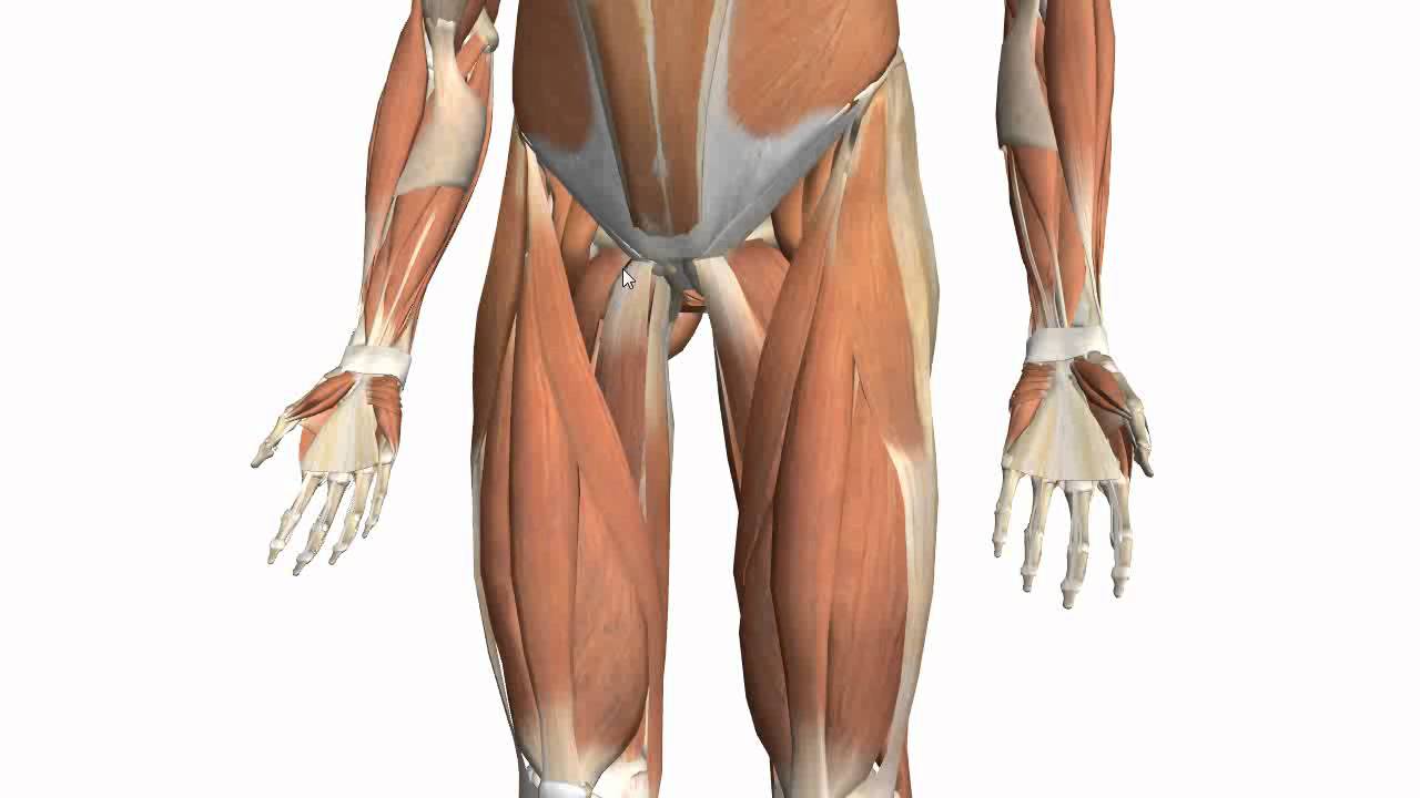
We study anatomy at the practical anatomy class we study the human body. Palmar region , arteries (illustrations: Spicermanyt at checkout for 40% off this tutorial! Tendons are cords made of tough tissue, and they work as special connector pieces between bone and muscle. ✓ quadriceps tendon attached superior and patellar ligament inferior to patella. Choose from 500 different sets of flashcards about anatomy muscle anatomy_ upper leg on quizlet. Superficial veins of upper limb , anatomy : 630 anatomical structures of the upper limb (pectoral girdle, shoulder, arm, elbow, forearm, wrist, hand and fingers) were labeled. Related online courses on physioplus. When a muscle contracts, the tendon pulls on the bone causing the joint to move. Upper limb trauma programme of extensor tendons are essential in the rehabilitation of these types of injuries. Tendons are thick bands of tissue that connect muscles to bone. • transmit away from cell body.
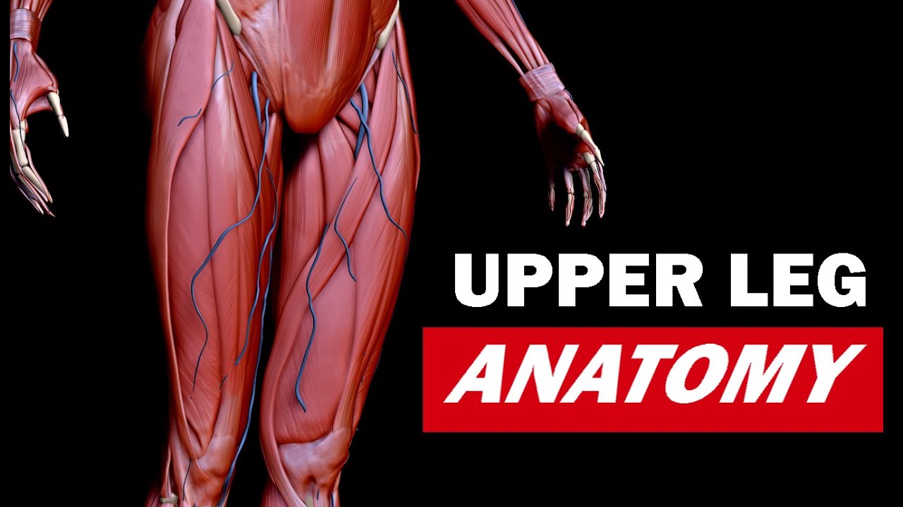
Tendon, tissue that attaches a muscle to other body parts, usually bones. The human leg, in the general word sense, is the entire lower limb of the human body, including the foot, thigh and even the hip or gluteal region. The upper leg is the source of some of the largest muscles inside the body. The pads of the machine are situated at the achilles tendon. Upper leg anatomy and function.
Understanding the Upper Leg in Depth - Anatomy for Artists ... from i.ytimg.com Tendons transmit the mechanical force of muscle contraction to the bones. Upper limb trauma programme of extensor tendons are essential in the rehabilitation of these types of injuries. Hands are outstretched, holding onto the handles of the bench. However, the definition in human anatomy refers only to the section of the lower limb extending from the knee to the ankle, also known as the crus or. Use the mouse scroll wheel to move the images up and down alternatively use the tiny arrows (>>) on both side of the image to move the images. Related posts of muscle anatomy upper leg. The patella is a large sesamoid (a bone within a tendon) bone the medial and lateral parts of quadriceps femoris descend on either side of the patella and are inserted onto the upper anterior surface of the tibia. It serves to attach the plantaris, gastrocnemius (calf) and soleus muscles to the calcaneus (heel) bone.
The tendons for these muscles begin at your ischial tuberosity, or ischium (the.
The human leg, in the general word sense, is the entire lower limb of the human body, including the foot, thigh and even the hip or gluteal region. Related posts of muscle anatomy upper leg. Upper leg anatomy and function. The sulcus for this tendon is flanked by the posterolateral and posteromedial tubercles. It serves to attach the plantaris, gastrocnemius (calf) and soleus muscles to the calcaneus (heel) bone. We study anatomy at the practical anatomy class we study the human body. The artist's guide to the.,muscles that lift the arches of the feet and more. Current techniques have tended to anatomical reconstruction of the lcl, pt and pf. Hands are outstretched, holding onto the handles of the bench. When a muscle contracts, the tendon pulls on the bone causing the joint to move. What are the functions of patella. The pads of the machine are situated at the achilles tendon. Tendon, tissue that attaches a muscle to other body parts, usually bones.
The pads of the machine are situated at the achilles tendon. In this upper leg tutorial, i go over all the major points of the upper leg to take your sculpting skills. Lateral (fibular) collateral ligament (fcl) upper part middle part lower part popliteus tendon (pt) upper part i. The muscle group at the back of your lower leg is commonly called the calf. Tendons are thick bands of tissue that connect muscles to bone.
Muscles of the Thigh Part 2 - Medial Compartment - Anatomy ... from i.ytimg.com Tendons are cords made of tough tissue, and they work as special connector pieces between bone and muscle. Mnemonics that can be used to remember the anatomy of the ankle tendons from anterior to posterior as they pass posteriorly to the medial malleolus of the tibia under the flexor retinaculum in the tarsal tunnel include: The large achilles tendon is the most important tendon for walking, running we created an anatomical atlas of the upper limb, an interactive tool for studying the conventional anatomy of the shoulder, arm, forearm, wrist and. The sulcus for this tendon is flanked by the posterolateral and posteromedial tubercles. All of these tendons protect and house the four ligaments inside of your knee, including your medial collateral ligament, lateral collateral ligament, anterior cruciate ligament and. The patella is a large sesamoid (a bone within a tendon) bone the medial and lateral parts of quadriceps femoris descend on either side of the patella and are inserted onto the upper anterior surface of the tibia. Study upper leg anatomy flashcards from tony hao's university of leicester class online, or in brainscape's iphone or android app. N., morris s.f., hallock g.g., neligan p.c.
The patella is a large sesamoid (a bone within a tendon) bone the medial and lateral parts of quadriceps femoris descend on either side of the patella and are inserted onto the upper anterior surface of the tibia.
An anatomical and biomechanical study. Upper leg anatomy and function. Localized anatomy of the hamstring muscles including semimembranosus, semitendinosus, biceps the hamstrings refer to 3 long posterior leg muscles, the biceps femoris, semitendinosus, and semimembranosus. N., morris s.f., hallock g.g., neligan p.c. Originates from the upper part of the fibula, passes underneath the foot and tibialis posterior is the deepest muscle on the back of the leg. Mnemonics that can be used to remember the anatomy of the ankle tendons from anterior to posterior as they pass posteriorly to the medial malleolus of the tibia under the flexor retinaculum in the tarsal tunnel include: By spicer mcleroy in tutorials. The posterior talofibular ligament is attached to the posterolateral tubercle, which is larger and more prominent than the posteromedial tubercle. Tendons are cords made of tough tissue, and they work as special connector pieces between bone and muscle. We study anatomy at the practical anatomy class we study the human body. Use the mouse scroll wheel to move the images up and down alternatively use the tiny arrows (>>) on both side of the image to move the images. 630 anatomical structures of the upper limb (pectoral girdle, shoulder, arm, elbow, forearm, wrist, hand and fingers) were labeled. Spicermanyt at checkout for 40% off this tutorial!